


What Happens During a Diabetic Eye Exam
If you have diabetes, a diabetic eye exam is one of the most important steps you can take to protect your vision. Diabetes can damage the small blood vessels in the retina, sometimes without early symptoms. A thorough diabetic eye exam looks for these changes before they affect your daily life.
At Paramount Eye Care, our optometry team uses detailed imaging and clinical testing to check for diabetic eye disease and other concerns that can impact long-term eye health. Here is what you can expect during a diabetic eye exam and why each step matters.
Why Diabetic Eye Exams Are Different From a Standard Eye Exam
A routine eye exam checks your prescription and overall eye health. A diabetic eye exam includes those basics, but it puts extra focus on the retina and the blood vessels at the back of the eye. The goal is to detect diabetic retinopathy, diabetic macular edema, and other diabetes-related changes early, when treatment and monitoring can be most effective.
Your provider may also look for related issues such as cataracts or glaucoma, which can occur more often in people with diabetes.
Step 1 - Health History and Diabetes Overview
Your visit usually starts with questions about your health and diabetes management. This helps your eye doctor understand your risk factors and decide which tests are most appropriate. Expect to discuss your A1C level, how long you have had diabetes, medications, blood pressure, and any vision changes such as blur, floaters, or trouble seeing at night.
Even if your vision feels stable, this step matters because diabetic eye disease can progress quietly.
Step 2 - Vision Testing and Eye Pressure Check
Next, you will complete vision testing to measure how clearly you see at distance and near. If you wear glasses or contacts, your prescription may be checked. Your provider may also measure eye pressure, since people with diabetes can have a higher risk of glaucoma.
These tests support a full picture of eye function and help separate prescription-related blur from retinal changes.
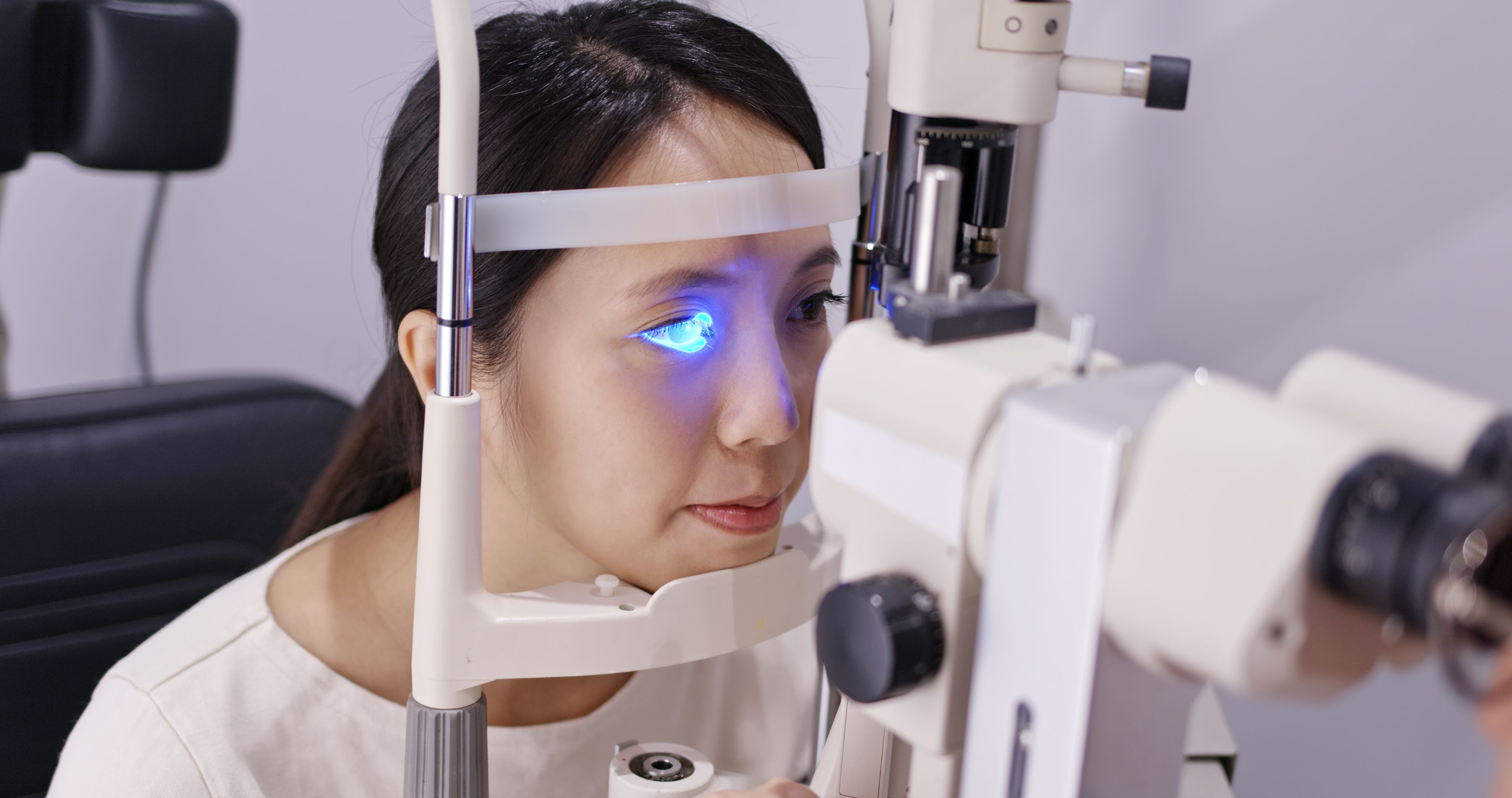
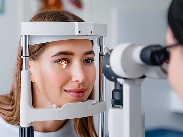
Step 3 - Retinal Evaluation with Dilation and Imaging
A key part of a diabetic eye exam is evaluating the retina. This will include pupil dilation, which allows your doctor to view the back of the eye in detail. After dilation, you may notice light sensitivity and blurry near vision for a few hours.
In addition to dilation, your appointment may include advanced retinal imaging. This can document the retina and help detect subtle swelling or vessel changes that are easy to miss early on.
One of the main benefits of combining clinical evaluation with imaging is improved tracking from year to year, especially if you have had diabetes for many years.
What Your Eye Doctor is Checking For
During a diabetic eye exam, your optometrist is looking for signs that diabetes is affecting the retina and surrounding structures, including:
- Diabetic retinopathy (leaking or damaged blood vessels)
- Diabetic macular edema (swelling in the central retina)
- Poor circulation or new abnormal blood vessel growth
- Retinal bleeding, fluid, or fatty deposits
- Cataract changes or increased glaucoma risk
Next Steps After Your Diabetic Eye Exam
After testing, your doctor will explain the findings and recommend next steps. If your eyes are healthy, you may simply need annual diabetic eye exams. If changes are found, your provider may recommend more frequent monitoring or a referral to a retina specialist depending on severity.
If you have diabetes, schedule your diabetic eye exam with Paramount Eye Care. Our office is in Lucas, Texas. Call 469-949-2020 to book an appointment today.

Contact Info
Hours of Operation
- Monday 9:00am - 6:00pm
- Tuesday 9:00am - 6:00pm
- Wednesday Closed
- Thursday 9:00am - 6:00pm
- Friday 9:00am - 5:00pm
- *Saturday 9:00am - 3:00pm
- Sunday Closed
- *Open 2 Saturdays per month, call for availability
© 2026 Paramount Eye Care and Eyewear. All rights Reserved. Accessibility Statement - Privacy Policy - Terms and Conditions - Sitemap
Powered by: